×
Network disruptions
We have been experiencing disruptions on our local network which has affected the stability of these web pages.
We have been working with IT support team to get this fixed as a matter of urgency and apologise for any inconvenience.
PDB Information
| PDB | 1SBN |
| Method | X-RAY DIFFRACTION |
| Host Organism | Escherichia coli |
| Gene Source | Bacillus subtilis |
| Primary Citation |
Refined crystal structures of subtilisin novo in complex with wild-type and two mutant eglins. Comparison with other serine proteinase inhibitor complexes.
Heinz, D.W., Priestle, J.P., Rahuel, J., Wilson, K.S., Grutter, M.G.
J.Mol.Biol.
|
| Header | Complex(Proteinase/Inhibitor) |
| Released | 1991-12-20 |
| Resolution | 2.100 |
| CATH Insert Date | 05 Mar, 2006 |
PDB Images (5)
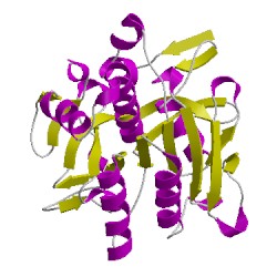
1sbnE00
CATH Domain 1sbnE00
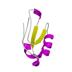
1sbnI00
CATH Domain 1sbnI00
PDB Chains (2)
| Chain ID | Date inserted into CATH | CATH Status |
| E | 05 Mar, 2006 | Chopped
|
| I | 05 Mar, 2006 | Chopped
|
CATH Domains (2)
UniProtKB Entries (2)
| Accession |
Gene ID |
Taxon |
Description |
| P01051 |
ICIC_HIRME |
Hirudo medicinalis |
Eglin C |
| P00782 |
SUBT_BACAM |
Bacillus amyloliquefaciens |
Subtilisin BPN' |