×
Network disruptions
We have been experiencing disruptions on our local network which has affected the stability of these web pages.
We have been working with IT support team to get this fixed as a matter of urgency and apologise for any inconvenience.
PDB Information
| PDB | 5YLA |
| Method | X-RAY DIFFRACTION |
| Host Organism | Escherichia coli |
| Gene Source | Acinetobacter baumannii |
| Primary Citation |
Crystal structure of a dimeric peptidyl-tRNA hydrolase from Acinetobacter baumannii at 1.67 A resolution
Singh, P.K., Sharma, P., Kaur, P., Sharma, S., Singh, T.P.
To Be Published
|
| Header | Hydrolase |
| Released | 2017-10-17 |
| Resolution | 1.680 |
| CATH Insert Date | 14 Nov, 2017 |
PDB Images (5)
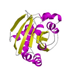
5ylaA00
CATH Domain 5ylaA00
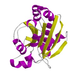
5ylaB00
CATH Domain 5ylaB00
PDB Chains (2)
| Chain ID | Date inserted into CATH | CATH Status |
| A | 14 Nov, 2017 | Chopped
|
| B | 14 Nov, 2017 | Chopped
|
CATH Domains (2)
UniProtKB Entries (2)
| Accession |
Gene ID |
Taxon |
Description |
| D0C9L6 |
D0C9L6_ACIB2 |
Acinetobacter baumannii ATCC 19606 = CIP 70.34 = JCM 6841 |
Peptidyl-tRNA hydrolase |
| D0C9L6 |
D0C9L6_ACIB2 |
Acinetobacter baumannii ATCC 19606 = CIP 70.34 = JCM 6841 |
Peptidyl-tRNA hydrolase |