×
Network disruptions
We have been experiencing disruptions on our local network which has affected the stability of these web pages.
We have been working with IT support team to get this fixed as a matter of urgency and apologise for any inconvenience.
PDB Information
| PDB | 5O0P |
| Method | X-RAY DIFFRACTION |
| Host Organism | Homo sapiens |
| Gene Source | Mus musculus |
| Primary Citation |
Nogo Receptor crystal structures with a native disulfide pattern suggest a novel mode of self-interaction.
Pronker, M.F., Tas, R.P., Vlieg, H.C., Janssen, B.J.C.
Acta Crystallogr D Struct Biol
|
| Header | Signaling Protein |
| Released | 2017-05-16 |
| Resolution | 2.000 |
| CATH Insert Date | 14 Nov, 2017 |
PDB Images (5)
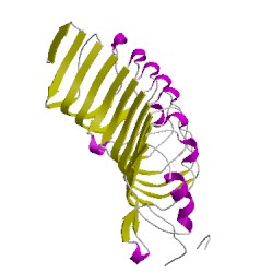
5o0pA00
CATH Domain 5o0pA00
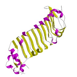
5o0pB00
CATH Domain 5o0pB00
PDB Chains (2)
| Chain ID | Date inserted into CATH | CATH Status |
| A | 14 Nov, 2017 | Chopped
|
| B | 14 Nov, 2017 | Chopped
|
CATH Domains (2)
UniProtKB Entries (2)
| Accession |
Gene ID |
Taxon |
Description |
| Q99PI8 |
RTN4R_MOUSE |
Mus musculus |
Reticulon-4 receptor |
| Q99PI8 |
RTN4R_MOUSE |
Mus musculus |
Reticulon-4 receptor |