×
Network disruptions
We have been experiencing disruptions on our local network which has affected the stability of these web pages.
We have been working with IT support team to get this fixed as a matter of urgency and apologise for any inconvenience.
PDB Information
| PDB | 3HRW |
| Method | X-RAY DIFFRACTION |
| Host Organism | |
| Gene Source | |
| Primary Citation |
Crystal structure of hemoglobin from mouse (Mus musculus)at 2.8
Sundaresan, S.S., Ramesh, P., Ponnuswamy, M.N.
To be Published
|
| Header | Oxygen Transport |
| Released | 2009-06-10 |
| Resolution | 2.800 |
| CATH Insert Date | 16 Aug, 2009 |
PDB Images (9)
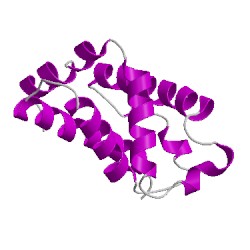
3hrwA00
CATH Domain 3hrwA00
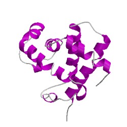
3hrwB00
CATH Domain 3hrwB00
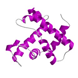
3hrwC00
CATH Domain 3hrwC00
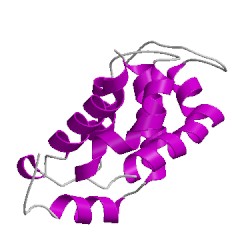
3hrwD00
CATH Domain 3hrwD00
PDB Chains (4)
CATH Domains (4)
UniProtKB Entries (4)
| Accession |
Gene ID |
Taxon |
Description |
| P01942 |
HBA_MOUSE |
Mus musculus |
Hemoglobin subunit alpha |
| P01942 |
HBA_MOUSE |
Mus musculus |
Hemoglobin subunit alpha |
| P02088 |
HBB1_MOUSE |
Mus musculus |
Hemoglobin subunit beta-1 |
| P02088 |
HBB1_MOUSE |
Mus musculus |
Hemoglobin subunit beta-1 |