×
Network disruptions
We have been experiencing disruptions on our local network which has affected the stability of these web pages.
We have been working with IT support team to get this fixed as a matter of urgency and apologise for any inconvenience.
PDB Information
| PDB | 3FMF |
| Method | X-RAY DIFFRACTION |
| Host Organism | Escherichia coli |
| Gene Source | Mycobacterium tuberculosis |
| Primary Citation |
Structural characterization of the Mycobacterium tuberculosis biotin biosynthesis enzymes 7,8-diaminopelargonic acid synthase and dethiobiotin synthetase .
Dey, S., Lane, J.M., Lee, R.E., Rubin, E.J., Sacchettini, J.C.
Biochemistry
|
| Header | Ligase |
| Released | 2008-12-21 |
| Resolution | 2.050 |
| CATH Insert Date | 16 Aug, 2009 |
PDB Images (9)
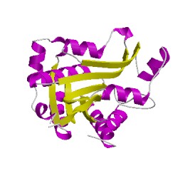
3fmfA00
CATH Domain 3fmfA00
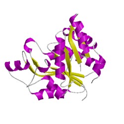
3fmfB00
CATH Domain 3fmfB00
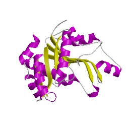
3fmfC00
CATH Domain 3fmfC00
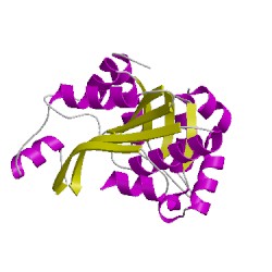
3fmfD00
CATH Domain 3fmfD00
PDB Chains (4)
CATH Domains (4)
UniProtKB Entries (4)
| Accession |
Gene ID |
Taxon |
Description |
| P9WPQ5 |
BIOD_MYCTU |
Mycobacterium tuberculosis H37Rv |
ATP-dependent dethiobiotin synthetase BioD |
| P9WPQ5 |
BIOD_MYCTU |
Mycobacterium tuberculosis H37Rv |
ATP-dependent dethiobiotin synthetase BioD |
| P9WPQ5 |
BIOD_MYCTU |
Mycobacterium tuberculosis H37Rv |
ATP-dependent dethiobiotin synthetase BioD |
| P9WPQ5 |
BIOD_MYCTU |
Mycobacterium tuberculosis H37Rv |
ATP-dependent dethiobiotin synthetase BioD |