×
Network disruptions
We have been experiencing disruptions on our local network which has affected the stability of these web pages.
We have been working with IT support team to get this fixed as a matter of urgency and apologise for any inconvenience.
PDB Information
| PDB | 3EQO |
| Method | X-RAY DIFFRACTION |
| Host Organism | Pichia pastoris |
| Gene Source | Phanerochaete chrysosporium |
| Primary Citation |
Crystal structure of glycoside hydrolase family 55 beta -1,3-glucanase from the basidiomycete Phanerochaete chrysosporium
Ishida, T., Fushinobu, S., Kawai, R., Kitaoka, M., Igarashi, K., Samejima, M.
J.Biol.Chem.
|
| Header | Hydrolase |
| Released | 2008-10-01 |
| Resolution | 2.250 |
| CATH Insert Date | 05 Feb, 2009 |
PDB Images (7)
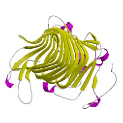
3eqoA01
CATH Domain 3eqoA01
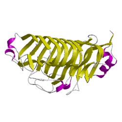
3eqoA02
CATH Domain 3eqoA02
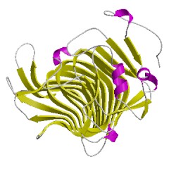
3eqoB01
CATH Domain 3eqoB01
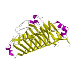
3eqoB02
CATH Domain 3eqoB02
PDB Chains (2)
| Chain ID | Date inserted into CATH | CATH Status |
| A | 05 Feb, 2009 | Chopped
|
| B | 05 Feb, 2009 | Chopped
|
CATH Domains (4)
UniProtKB Entries (2)
| Accession |
Gene ID |
Taxon |
Description |
| Q2Z1W1 |
Q2Z1W1_PHACH |
Phanerochaete chrysosporium |
Glucan 1,3-beta-glucosidase |
| Q2Z1W1 |
Q2Z1W1_PHACH |
Phanerochaete chrysosporium |
Glucan 1,3-beta-glucosidase |