×
Network disruptions
We have been experiencing disruptions on our local network which has affected the stability of these web pages.
We have been working with IT support team to get this fixed as a matter of urgency and apologise for any inconvenience.
PDB Information
| PDB | 3DGT |
| Method | X-RAY DIFFRACTION |
| Host Organism | Escherichia coli |
| Gene Source | Streptomyces sioyaensis |
| Primary Citation |
The 1.5 A structure of endo-1,3-beta-glucanase from Streptomyces sioyaensis: evolution of the active-site structure for 1,3-beta-glucan-binding specificity and hydrolysis
Hong, T.-Y., Hsiao, Y.-Y., Meng, M., Li, T.T.
Acta Crystallogr.,Sect.D
|
| Header | Hydrolase |
| Released | 2008-06-16 |
| Resolution | 1.500 |
| CATH Insert Date | 04 Sep, 2008 |
PDB Images (3)
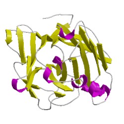
3dgtA00
CATH Domain 3dgtA00
PDB Chains (1)
| Chain ID | Date inserted into CATH | CATH Status |
| A | 04 Sep, 2008 | Chopped
|
CATH Domains (1)
UniProtKB Entries (1)
| Accession |
Gene ID |
Taxon |
Description |
| Q9L816 |
Q9L816_9ACTN |
Streptomyces sioyaensis |
Endo-1,3-beta-glucanase |