×
Network disruptions
We have been experiencing disruptions on our local network which has affected the stability of these web pages.
We have been working with IT support team to get this fixed as a matter of urgency and apologise for any inconvenience.
PDB Information
| PDB | 1QBI |
| Method | X-RAY DIFFRACTION |
| Host Organism | Escherichia coli |
| Gene Source | Acinetobacter calcoaceticus |
| Primary Citation |
The 1.7 A crystal structure of the apo form of the soluble quinoprotein glucose dehydrogenase from Acinetobacter calcoaceticus reveals a novel internal conserved sequence repeat.
Oubrie, A., Rozeboom, H.J., Kalk, K.H., Duine, J.A., Dijkstra, B.W.
J.Mol.Biol.
|
| Header | Oxidoreductase |
| Released | 1999-04-22 |
| Resolution | 1.720 |
| CATH Insert Date | 05 Mar, 2006 |
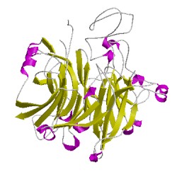
PDB Images (5)

1qbiA00
CATH Domain 1qbiA00
1qbiB00
CATH Domain 1qbiB00
PDB Chains (2)
| Chain ID | Date inserted into CATH | CATH Status |
| A | 05 Mar, 2006 | Chopped
|
| B | 05 Mar, 2006 | Chopped
|
CATH Domains (2)
UniProtKB Entries (2)
| Accession |
Gene ID |
Taxon |
Description |
| P13650 |
DHGB_ACICA |
Acinetobacter calcoaceticus |
Quinoprotein glucose dehydrogenase B |
| P13650 |
DHGB_ACICA |
Acinetobacter calcoaceticus |
Quinoprotein glucose dehydrogenase B |