×
Network disruptions
We have been experiencing disruptions on our local network which has affected the stability of these web pages.
We have been working with IT support team to get this fixed as a matter of urgency and apologise for any inconvenience.
PDB Information
| PDB | 1PFR |
| Method | X-RAY DIFFRACTION |
| Host Organism | Escherichia coli |
| Gene Source | Escherichia coli |
| Primary Citation |
Crystal structure of reduced protein R2 of ribonucleotide reductase: the structural basis for oxygen activation at a dinuclear iron site.
Logan, D.T., Su, X.D., Aberg, A., Regnstrom, K., Hajdu, J., Eklund, H., Nordlund, P.
Structure
|
| Header | Reductase |
| Released | 1996-12-03 |
| Resolution | 2.200 |
| CATH Insert Date | 05 Mar, 2006 |
PDB Images (5)
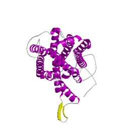
1pfrA00
CATH Domain 1pfrA00
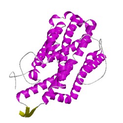
1pfrB00
CATH Domain 1pfrB00
PDB Chains (2)
| Chain ID | Date inserted into CATH | CATH Status |
| A | 05 Mar, 2006 | Chopped
|
| B | 05 Mar, 2006 | Chopped
|
CATH Domains (2)
UniProtKB Entries (2)
| Accession |
Gene ID |
Taxon |
Description |
| P69924 |
RIR2_ECOLI |
Escherichia coli K-12 |
Ribonucleoside-diphosphate reductase 1 subunit beta |
| P69924 |
RIR2_ECOLI |
Escherichia coli K-12 |
Ribonucleoside-diphosphate reductase 1 subunit beta |