×
Network disruptions
We have been experiencing disruptions on our local network which has affected the stability of these web pages.
We have been working with IT support team to get this fixed as a matter of urgency and apologise for any inconvenience.
PDB Information
| PDB | 5AXD |
| Method | X-RAY DIFFRACTION |
| Host Organism | Escherichia coli |
| Gene Source | Mus musculus |
| Primary Citation |
Crystal structure of mouse SAHH complexed with ribavirin
Kusakabe, Y., Ishihara, M., Tanaka, N.
To Be Published
|
| Header | Hydrolase |
| Released | 2015-07-24 |
| Resolution | 1.600 |
| CATH Insert Date | 07 Aug, 2016 |
PDB Images (7)
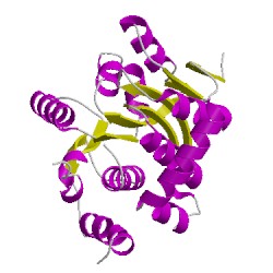
5axdA01
CATH Domain 5axdA01
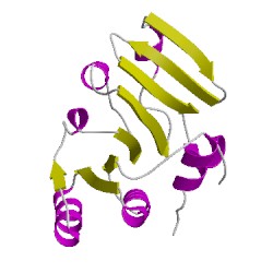
5axdA02
CATH Domain 5axdA02
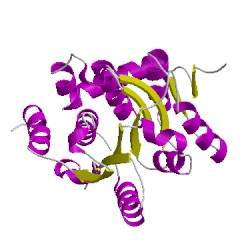
5axdC01
CATH Domain 5axdC01
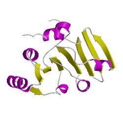
5axdC02
CATH Domain 5axdC02
PDB Chains (2)
| Chain ID | Date inserted into CATH | CATH Status |
| A | 08 Aug, 2016 | Chopped
|
| C | 08 Aug, 2016 | Chopped
|
CATH Domains (4)
UniProtKB Entries (2)
| Accession |
Gene ID |
Taxon |
Description |
| P50247 |
SAHH_MOUSE |
Mus musculus |
Adenosylhomocysteinase |
| P50247 |
SAHH_MOUSE |
Mus musculus |
Adenosylhomocysteinase |