×
Network disruptions
We have been experiencing disruptions on our local network which has affected the stability of these web pages.
We have been working with IT support team to get this fixed as a matter of urgency and apologise for any inconvenience.
PDB Information
| PDB | 4B1W |
| Method | X-RAY DIFFRACTION |
| Host Organism | |
| Gene Source | |
| Primary Citation |
Structures of the Phactr1 RPEL domain and RPEL motif complexes with G-actin reveal the molecular basis for actin binding cooperativity.
Mouilleron, S., Wiezlak, M., O'Reilly, N., Treisman, R., McDonald, N.Q.
Structure
|
| Header | Structural Protein |
| Released | 2012-07-12 |
| Resolution | 1.950 |
| CATH Insert Date | 18 Aug, 2013 |
PDB Images (6)
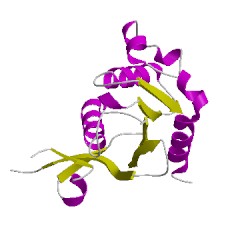
4b1wB01
CATH Domain 4b1wB01
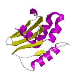
4b1wB02
CATH Domain 4b1wB02
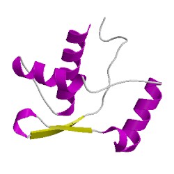
4b1wB03
CATH Domain 4b1wB03
PDB Chains (2)
| Chain ID | Date inserted into CATH | CATH Status |
| B | 19 Aug, 2013 | Chopped
|
| M | 19 Aug, 2013 | Rejected
|
CATH Domains (3)
UniProtKB Entries (2)
| Accession |
Gene ID |
Taxon |
Description |
| Q2M3X8 |
PHAR1_MOUSE |
Mus musculus |
Phosphatase and actin regulator 1 |
| P68135 |
ACTS_RABIT |
Oryctolagus cuniculus |
Actin, alpha skeletal muscle |