×
Network disruptions
We have been experiencing disruptions on our local network which has affected the stability of these web pages.
We have been working with IT support team to get this fixed as a matter of urgency and apologise for any inconvenience.
PDB Information
| PDB | 3D02 |
| Method | X-RAY DIFFRACTION |
| Host Organism | Escherichia coli |
| Gene Source | Klebsiella pneumoniae subsp. pneumoniae |
| Primary Citation |
Crystal structure of periplasmic sugar-binding protein (YP_001338366.1) from Klebsiella pneumoniae subsp. pneumoniae MGH 78578 at 1.30 A resolution
Joint Center for Structural Genomics (JCSG)
To be published
|
| Header | Sugar Binding Protein |
| Released | 2008-04-30 |
| Resolution | 1.300 |
| CATH Insert Date | 23 Jul, 2008 |
PDB Images (4)
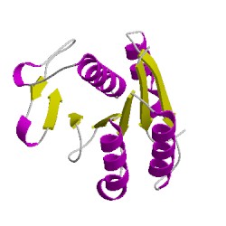
3d02A01
CATH Domain 3d02A01
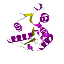
3d02A02
CATH Domain 3d02A02
PDB Chains (1)
| Chain ID | Date inserted into CATH | CATH Status |
| A | 24 Jul, 2008 | Chopped
|
CATH Domains (2)
UniProtKB Entries (1)
| Accession |
Gene ID |
Taxon |
Description |
| A6THR5 |
A6THR5_KLEP7 |
Klebsiella pneumoniae subsp. pneumoniae MGH 78578 |
Putative LACI-type transcriptional regulator |