×
Network disruptions
We have been experiencing disruptions on our local network which has affected the stability of these web pages.
We have been working with IT support team to get this fixed as a matter of urgency and apologise for any inconvenience.
PDB Information
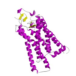
| PDB | 3C9L |
| Method | X-RAY DIFFRACTION |
| Host Organism | |
| Gene Source | |
| Primary Citation |
Alternative models for two crystal structures of bovine rhodopsin.
Stenkamp, R.E.
Acta Crystallogr.,Sect.D
|
| Header | Signaling Protein |
| Released | 2008-02-16 |
| Resolution | 2.650 |
| CATH Insert Date | 07 Aug, 2008 |
PDB Images (3)
3c9lA00
CATH Domain 3c9lA00
PDB Chains (1)
| Chain ID | Date inserted into CATH | CATH Status |
| A | 07 Aug, 2008 | Chopped
|
CATH Domains (1)
UniProtKB Entries (1)
| Accession |
Gene ID |
Taxon |
Description |
| P02699 |
OPSD_BOVIN |
Bos taurus |
Rhodopsin |