×
Network disruptions
We have been experiencing disruptions on our local network which has affected the stability of these web pages.
We have been working with IT support team to get this fixed as a matter of urgency and apologise for any inconvenience.
PDB Information
| PDB | 1VED |
| Method | X-RAY DIFFRACTION |
| Host Organism | |
| Gene Source | |
| Primary Citation |
The crystal structure of the orthorhombic form of hen egg white lysozyme at 1.9 angstroms resolution in space
Aibara, S., Suzuki, A., Kidera, A., Shibata, K., Yamane, T., DeLucas, L.J., Hirose, M.
To be Published
|
| Header | Hydrolase |
| Released | 2004-03-30 |
| Resolution | 1.900 |
| CATH Insert Date | 05 Mar, 2006 |
PDB Images (3)
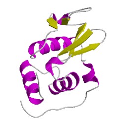
1vedA00
CATH Domain 1vedA00
PDB Chains (1)
| Chain ID | Date inserted into CATH | CATH Status |
| A | 05 Mar, 2006 | Chopped
|
CATH Domains (1)
UniProtKB Entries (1)
| Accession |
Gene ID |
Taxon |
Description |
| P00698 |
LYSC_CHICK |
Gallus gallus |
Lysozyme C |