×
Network disruptions
We have been experiencing disruptions on our local network which has affected the stability of these web pages.
We have been working with IT support team to get this fixed as a matter of urgency and apologise for any inconvenience.
PDB Information
| PDB | 1P9L |
| Method | X-RAY DIFFRACTION |
| Host Organism | Escherichia coli BL21(DE3) |
| Gene Source | Mycobacterium tuberculosis |
| Primary Citation |
The three-dimensional structures of the Mycobacterium tuberculosis dihydrodipicolinate reductase-NADH-2,6-PDC and -NADPH-2,6-PDC complexes. Structural and mutagenic analysis of relaxed nucleotide specificity.
Cirilli, M., Zheng, R., Scapin, G., Blanchard, J.S.
Biochemistry
|
| Header | Oxidoreductase |
| Released | 2003-05-12 |
| Resolution | 2.300 |
| CATH Insert Date | 05 Mar, 2006 |
PDB Images (7)
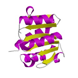
1p9lA01
CATH Domain 1p9lA01
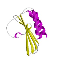
1p9lA02
CATH Domain 1p9lA02
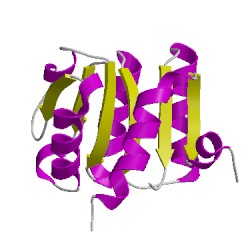
1p9lB01
CATH Domain 1p9lB01
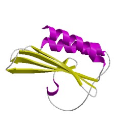
1p9lB02
CATH Domain 1p9lB02
PDB Chains (2)
| Chain ID | Date inserted into CATH | CATH Status |
| A | 05 Mar, 2006 | Chopped
|
| B | 05 Mar, 2006 | Chopped
|
CATH Domains (4)
UniProtKB Entries (2)
| Accession |
Gene ID |
Taxon |
Description |
| P9WP23 |
DAPB_MYCTU |
Mycobacterium tuberculosis H37Rv |
4-hydroxy-tetrahydrodipicolinate reductase |
| P9WP23 |
DAPB_MYCTU |
Mycobacterium tuberculosis H37Rv |
4-hydroxy-tetrahydrodipicolinate reductase |