×
Network disruptions
We have been experiencing disruptions on our local network which has affected the stability of these web pages.
We have been working with IT support team to get this fixed as a matter of urgency and apologise for any inconvenience.
PDB Information
| PDB | 1OGF |
| Method | X-RAY DIFFRACTION |
| Host Organism | |
| Gene Source | |
| Primary Citation |
Crystal Structures of Rbsd Leading to the Identification of Cytoplasmic Sugar-Binding Proteins with a Novel Folding Architecture
Kim, M.-S., Shin, J., Lee, W., Lee, H.-S., Oh, B.-H.
J.Biol.Chem.
|
| Header | Transport |
| Released | 2003-04-30 |
| Resolution | 2.300 |
| CATH Insert Date | 05 Mar, 2006 |
PDB Images (11)
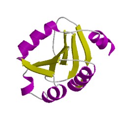
1ogfA01
CATH Domain 1ogfA01
1ogfB01
CATH Domain 1ogfB01
1ogfC01
CATH Domain 1ogfC01
1ogfD01
CATH Domain 1ogfD01
1ogfE01
CATH Domain 1ogfE01
PDB Chains (5)
CATH Domains (5)
UniProtKB Entries (5)
| Accession |
Gene ID |
Taxon |
Description |
| P36946 |
RBSD_BACSU |
Bacillus subtilis subsp. subtilis str. 168 |
D-ribose pyranase |
| P36946 |
RBSD_BACSU |
Bacillus subtilis subsp. subtilis str. 168 |
D-ribose pyranase |
| P36946 |
RBSD_BACSU |
Bacillus subtilis subsp. subtilis str. 168 |
D-ribose pyranase |
| P36946 |
RBSD_BACSU |
Bacillus subtilis subsp. subtilis str. 168 |
D-ribose pyranase |
| P36946 |
RBSD_BACSU |
Bacillus subtilis subsp. subtilis str. 168 |
D-ribose pyranase |