×
Network disruptions
We have been experiencing disruptions on our local network which has affected the stability of these web pages.
We have been working with IT support team to get this fixed as a matter of urgency and apologise for any inconvenience.
PDB Information
| PDB | 1K96 |
| Method | X-RAY DIFFRACTION |
| Host Organism | Escherichia coli |
| Gene Source | Homo sapiens |
| Primary Citation |
Crystal structures of S100A6 in the Ca(2+)-free and Ca(2+)-bound states: the calcium sensor mechanism of S100 proteins revealed at atomic resolution.
Otterbein, L.R., Kordowska, J., Witte-Hoffmann, C., Wang, C.L., Dominguez, R.
Structure
|
| Header | Signaling Protein |
| Released | 2001-10-26 |
| Resolution | 1.440 |
| CATH Insert Date | 05 Mar, 2006 |
PDB Images (3)
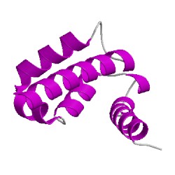
1k96A00
CATH Domain 1k96A00
PDB Chains (1)
| Chain ID | Date inserted into CATH | CATH Status |
| A | 05 Mar, 2006 | Chopped
|
CATH Domains (1)
UniProtKB Entries (1)
| Accession |
Gene ID |
Taxon |
Description |
| P06703 |
S10A6_HUMAN |
Homo sapiens |
Protein S100-A6 |