×
Network disruptions
We have been experiencing disruptions on our local network which has affected the stability of these web pages.
We have been working with IT support team to get this fixed as a matter of urgency and apologise for any inconvenience.
PDB Information
| PDB | 1I7X |
| Method | X-RAY DIFFRACTION |
| Host Organism | Escherichia coli |
| Gene Source | Mus musculus |
| Primary Citation |
The structure of the beta-catenin/E-cadherin complex and the molecular basis of diverse ligand recognition by beta-catenin.
Huber, A.H., Weis, W.I.
Cell(Cambridge,Mass.)
|
| Header | Cell Adhesion |
| Released | 2001-03-10 |
| Resolution | 3.000 |
| CATH Insert Date | 05 Mar, 2006 |
PDB Images (9)
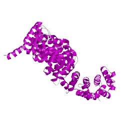
1i7xA00
CATH Domain 1i7xA00
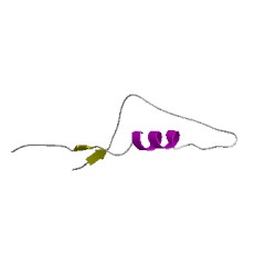
1i7xB00
CATH Domain 1i7xB00
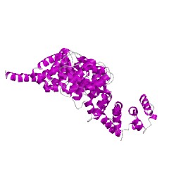
1i7xC00
CATH Domain 1i7xC00
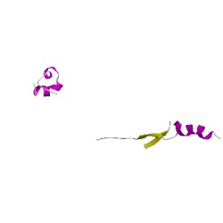
1i7xD00
CATH Domain 1i7xD00
PDB Chains (4)
CATH Domains (4)
UniProtKB Entries (4)
| Accession |
Gene ID |
Taxon |
Description |
| P09803 |
CADH1_MOUSE |
Mus musculus |
Cadherin-1 |
| Q02248 |
CTNB1_MOUSE |
Mus musculus |
Catenin beta-1 |
| P09803 |
CADH1_MOUSE |
Mus musculus |
Cadherin-1 |
| Q02248 |
CTNB1_MOUSE |
Mus musculus |
Catenin beta-1 |