×
Network disruptions
We have been experiencing disruptions on our local network which has affected the stability of these web pages.
We have been working with IT support team to get this fixed as a matter of urgency and apologise for any inconvenience.
PDB Information
| PDB | 1H8U |
| Method | X-RAY DIFFRACTION |
| Host Organism | |
| Gene Source | |
| Primary Citation |
Crystal Structure of the Eosinophil Major Basic Protein at 1.8A. An Atypical Lectin with a Paradigm Shift in Specificity
Swaminathan, G.J., Weaver, A.J., Loegering, D.A., Checkel, J.L., Leonidas, D.D., Gleich, G.J., Acharya, K.R.
J.Biol.Chem.
|
| Header | Lectin |
| Released | 2001-02-15 |
| Resolution | 1.800 |
| CATH Insert Date | 05 Mar, 2006 |
PDB Images (5)
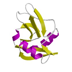
1h8uA00
CATH Domain 1h8uA00
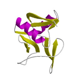
1h8uB00
CATH Domain 1h8uB00
PDB Chains (2)
| Chain ID | Date inserted into CATH | CATH Status |
| A | 05 Mar, 2006 | Chopped
|
| B | 05 Mar, 2006 | Chopped
|
CATH Domains (2)
UniProtKB Entries (2)
| Accession |
Gene ID |
Taxon |
Description |
| P13727 |
PRG2_HUMAN |
Homo sapiens |
Bone marrow proteoglycan |
| P13727 |
PRG2_HUMAN |
Homo sapiens |
Bone marrow proteoglycan |